

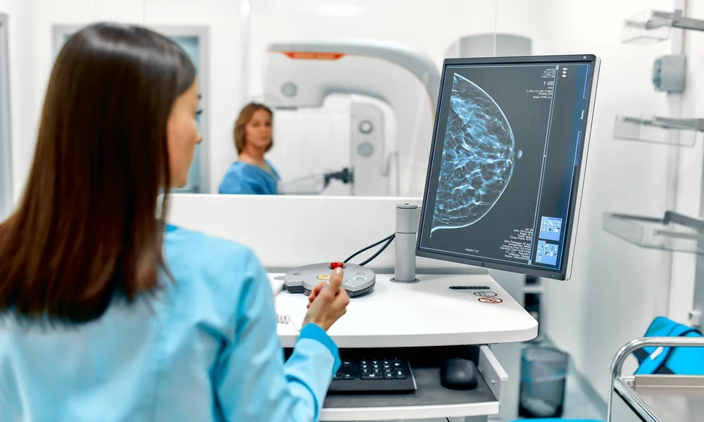
Breast cancer screening has evolved significantly over the past three decades, yet the journey from initial detection to definitive diagnosis remains complex for many women. While mammography serves as the cornerstone of screening programs worldwide, certain breast tissue types and suspicious findings require more sophisticated imaging approaches. Understanding when standard tests fall short-and what advanced options exist-empowers patients to make informed decisions about their diagnostic pathway.
The Limitations of Standard Mammography
Mammography has reduced breast cancer mortality by approximately 20% in screened populations, but it faces inherent challenges with dense breast tissue. Dense breasts contain more glandular and fibrous tissue relative to fatty tissue, creating a “white-on-white” effect where potential tumors and normal tissue appear similarly on X-ray images. Studies indicate that mammography sensitivity drops from roughly 87% in fatty breasts to 62% in extremely dense breasts.
This limitation affects nearly half of all women undergoing screening, particularly those under 50. When a mammogram reveals an area of concern in dense tissue-or when a palpable lump doesn’t show clearly on standard imaging-clinicians must turn to supplemental technologies that offer greater tissue differentiation.
When Ultrasound and Biopsy Guidance Fall Short
Ultrasound serves as the first-line supplemental tool for evaluating breast abnormalities, especially in younger women with dense tissue. It excels at distinguishing fluid-filled cysts from solid masses and guides many needle biopsies with real-time visualization. However, ultrasound has blind spots. Some lesions visible on mammography-particularly certain calcification patterns or architectural distortions-remain invisible on ultrasound, making biopsy targeting impossible through this method alone.
Stereotactic biopsy, which uses mammographic guidance and computer coordinates, addresses some of these cases but requires compression and positioning that can be uncomfortable and sometimes technically challenging. The procedure works well for calcifications but struggles with lesions deep in the breast or near the chest wall. For women with specific types of suspicious findings, neither ultrasound nor stereotactic approaches provide adequate visualization for confident tissue sampling.
Advanced Imaging for Complex Diagnostic Scenarios
Magnetic resonance imaging of the breast operates on fundamentally different principles than mammography or ultrasound. Rather than using X-rays or sound waves, MRI employs powerful magnets and radio waves to create detailed cross-sectional images of breast tissue. The technology detects differences in how various tissues respond to magnetic fields, particularly after injection of a contrast agent that highlights areas with increased blood flow-a hallmark of many cancers.
MRI demonstrates superior sensitivity for detecting breast cancer, particularly in high-risk patients and those with dense tissue. When a suspicious area appears on MRI but remains occult on other imaging modalities, specialized facilities can perform mri biopsy breast cancer procedures that use the same imaging technology to guide needle placement with submillimeter precision. This approach allows clinicians to sample lesions that would otherwise remain inaccessible, providing definitive pathology results without surgical intervention.
The Role of Specialized Medical Centers
Advanced diagnostic procedures require not only sophisticated equipment but also radiologists and technologists with specific training in breast imaging subspecialties. Multi-specialty hospitals with dedicated breast centers typically maintain the infrastructure necessary for the full range of diagnostic approaches, from routine screening through complex interventional procedures. Healthcare facilities in Abu Dhabi have expanded their imaging capabilities in recent years, reflecting growing awareness of the need for comprehensive diagnostic services in the region.
Access to advanced imaging becomes particularly important for international patients who travel to the UAE for specialized care. Coordinating multiple diagnostic steps-initial imaging, biopsy, pathology review, and treatment planning-within a single medical system streamlines what can otherwise become a fragmented process across multiple providers.
Patient Selection for Advanced Imaging
Not every patient requires MRI-based evaluation. Clinical guidelines reserve advanced imaging for specific scenarios: women with BRCA1 or BRCA2 genetic mutations, those with a lifetime breast cancer risk exceeding 20%, patients with a personal history of breast cancer undergoing surveillance, and cases where standard imaging yields inconclusive results. Overuse of highly sensitive imaging can lead to false positives and unnecessary interventions, making appropriate patient selection critical.
What Patients Should Know About Contrast Agents and Reactions
MRI breast imaging typically requires intravenous administration of gadolinium-based contrast agents to enhance visualization of suspicious areas. While generally well-tolerated, these substances can trigger reactions in sensitive individuals. Most reactions are mild-warmth, metallic taste, or nausea-but severe allergic responses occur in roughly 1 in 10,000 administrations.
Patients with known sensitivities to other medical compounds should inform their care team before any contrast-enhanced procedure. The broader issue of inactive ingredients in medical products causing unexpected reactions extends beyond imaging agents to medications and other healthcare materials, making comprehensive allergy histories essential for safe care delivery.
Interpreting Results and Next Steps
Biopsy results from advanced imaging-guided procedures follow the same pathology classification system used for all breast tissue samples. Results range from benign findings requiring no further action to atypical hyperplasia warranting increased surveillance, to various grades of carcinoma necessitating treatment. The precision of image-guided sampling reduces the likelihood of non-diagnostic results, but no technique achieves perfect accuracy.
When pathology reveals cancer, the same imaging that guided the biopsy often plays a role in treatment planning. MRI helps surgeons determine tumor extent, identify additional lesions, and assess the opposite breast. This information influences decisions about breast-conserving surgery versus mastectomy, and whether chemotherapy before surgery might shrink the tumor enough to allow less extensive removal.
Making Informed Decisions About Your Diagnostic Path
Women facing recommendations for advanced breast imaging should approach the situation with both the seriousness it deserves and an understanding that sophisticated evaluation tools exist precisely for complex cases. Questions to discuss with your care team include why standard approaches proved insufficient, whether your specific finding type is best evaluated with the proposed imaging method, what alternatives exist, and how results will inform treatment decisions.
The diagnostic journey from screening abnormality to definitive diagnosis involves multiple decision points, each informed by prior results and individual risk factors. While the process can feel overwhelming, modern imaging technology-when appropriately applied-provides answers with minimal invasiveness, allowing patients and clinicians to move forward with treatment planning based on accurate pathology rather than uncertainty.